Infrared Diagnostic Unit Of Mammary Gland is designed according to the different infrared absorption ratio of human bio-tissue. After the transillumination of the mammary tissue by infrared probe, signals will be photo-electrically transmitted and then processed in the computer, and the screen will show the pathological changes. The images processed help with quick and accurate diagnosis. Benign or malignant tumor, hyperplasia of mammary glands, breast cyst and other galactophore diseases can be identified according to the different shades and boundaries of the shadows, as well as the distribution of the vessels. The clinical application verifies that it is a quick, accurate, none-invasive diagnostic method, having special significance for detecting early breast cancer, and superior to the traditional examining methods such as the x-rays and the B-ultrasound.
The most advanced technologies in the relevant fields, such as electronics, computer and optics, are introduced into the making of the instrument. The private image data acquisition card supported by imported PCI bus, together with a private accelerating graphic card, presents lively color, abundant layers of the black and white image, and high contrast ratio. Windows 98 and windows XP in Chinese interface, plus the key press operation are convenient for doctor manipulation and at the same time, provide the operators with a safe and reliable system.
Instrument characteristics:
1.17-inch color perfectly flat display of high resolution produces high quality images.
2. Functions include two images or four images comparison in the same screen, double images comparison in dual-screens, case identification and diagnosis.
3. Rapid and convenient image acquisition methods include: Pedal, mouse, keyboard, etc.
4. A self-developed high sensitive double infrared probe, with its continuous adjustable brightness, strong tissue penetration and reliable quality, is able to examine different mammary gland tissues.
5. The highly sensitive camera enables high identification image taking in the darkroom.
6. Digital image card is used to ensure high clear and soft images of the galactophore.
7. The instrument is equipped with well-illustrated Chinese interface encoded in multi-pseudo color and possesses powerful image processing function which include: Gray image processing, image zooming in and zooming out, histogram, image enhancement, selective enlargement, negative photographic display, etc. It is also characterized by easy measurement of length, girth and area and convenient character label.
8. Galactophore examination report, term library and universality report templates can be obtained with speed.
9. It is equipped with practical database document management. Medical records and images can be stored in general format with no compressionloss and be ready for call. Management of medical records and images is available. The instrument has a storage capacity of over 100 thousand images and provides easy access to them.
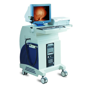
10. Available models include: Single screen handcart, dual-screen handcart and portable.
Technical specification:
1. Effective probe spectral wavelength range: 0.8~1.5μm
2. Adjustable probe output power range: 0.2~1W
3. Image resolution:>400TVL
4. Image processing function includes: 8 sets colors, system setting, image scaling, calibration setting, measurements of length, circumference and area, focus note addition, image stretching, two-value image processing, left and right separated screen and 32-pesudocolor.
Configuration standard:
1. Computer: Brand new computer
2. Display: 17 -inch color perfectly flat display or LCD
3. Camera: Resolution >420TVL
4. Printer: Color ink-jet printer of EPSON or HP
5. Strong infrared fiber probe
6. Luxurious handcar
|
|